Artificial Intelligence to Detect Papilledema from Ocular Fundus Photographs
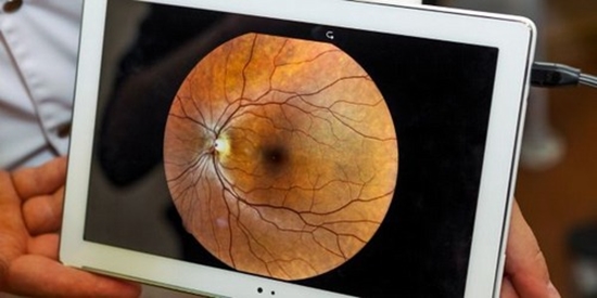
onophthalmologist physicians do not confidently perform direct ophthalmoscopy. The use of artificial intelligence to detect papilledema and other optic-disk abnormalities from fundus photographs has not been well studied.
We trained, validated, and externally tested a deep-learning system to classify optic disks as being normal or having papilledema or other abnormalities from 15,846 retrospectively collected ocular fundus photographs that had been obtained with pharmacologic pupillary dilation and various digital cameras in persons from multiple ethnic populations. Of these photographs, 14,341 from 19 sites in 11 countries were used for training and validation, and 1505 photographs from 5 other sites were used for external testing. Performance at classifying the optic-disk appearance was evaluated by calculating the area under the receiver-operating-characteristic curve (AUC), sensitivity, and specificity, as compared with a reference standard of clinical diagnoses by neuro-ophthalmologists.

ارسال نظر